Xray Vision Shoulders and Elbows — Taming the SRU Medical anatomy

Pin on Radiology
In order to identify pathology on shoulder plain films, an understanding of normal anatomy is essential. There are three main components of the shoulder radiography: the bones, the joints and soft tissue. The bones of the shoulder include the proximal humerus, the lateral clavicle, the ribs, and the scapula. The scapula is, in turn, subdivided.

shoulder xray oa3 DOCJOINTS//DR SUJIT JOS//Total joint replacements
Shoulder X-Ray. A shoulder X-ray uses radiation to take pictures of the bones and structures in your shoulder. Healthcare providers use a shoulder X-ray to diagnose conditions like broken bones, arthritis and dislocation. Shoulder X-rays are noninvasive and not painful. Contents Overview Test Details Results and Follow-Up.

Xray Vision Shoulders and Elbows — Taming the SRU Medical anatomy
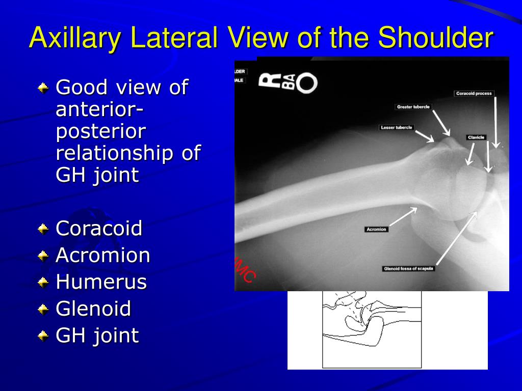
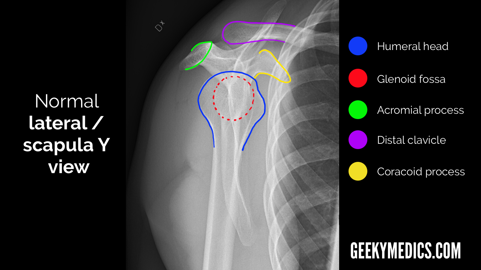
The lateral contour of the shoulder should be positioned in front of the film in a way that the longitudinal axis of the scapula continues parallel to the path of the rays. This view reveals: The horizontal centralization of the humerus head and socket. The osseous margins of the coraco-acromial arch and hence the supraspinatus outlet canal.
Shoulder XRay Right acromioclavicular joint dislocation radRounds
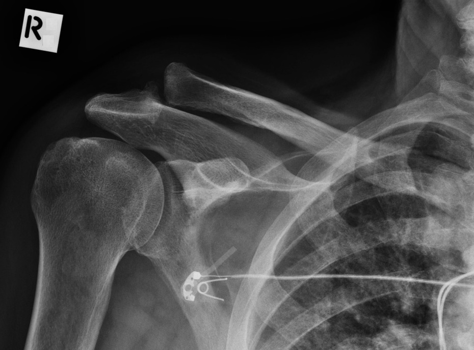
marked fragmentation, debris, and subluxation or dislo-cation of the humeral head. The changes can occur very rapidly often over the course of a few weeks and it often leads to the appearance of surgically amputated ends of bones61 (Fig. 21A and B). The differential diagnosis in-cludes infection and tumor.
PPT XRay Rounds (Plain) Radiographic Evaluation of the Shoulder
This projection is a true anterior-posterior (AP) view of the shoulder. The Grashey view involves angling the beam laterally or rotating the patient posteriorly(2). These adjustments remove the view of the overlap between the humerus and the glenoid. The removal allows better evaluation of joint congruity, humeral head subluxation, and the.
Shoulder Xray Century City Los Angeles, CA Commons Clinic
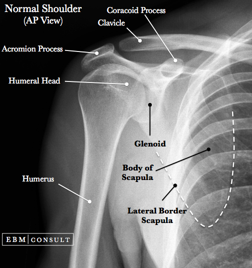
Citation, DOI, disclosures and article data. The shoulder AP view is a standard projection that makes up the two view shoulder series. The projection demonstrates the shoulder in its natural anatomical position allowing for adequate radiographic examination of the entire clavicle and scapula, as well as the glenohumeral, acromioclavicular and.
Anterior Shoulder Dislocation General Review
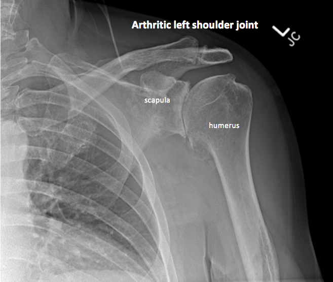
Macroscopic Functional Anatomy. The head and the glenoid fossa articulate in the shoulder joint (glenohumeral joint). Functionally, it is a ball-and-socket joint that enables movement in three degrees of freedom. The shoulder is the most mobile of the major joints. Its high mobility, together with its limited osseous embracement accounts for.
Shoulder Xray Interpretation Radiology Geeky Medics
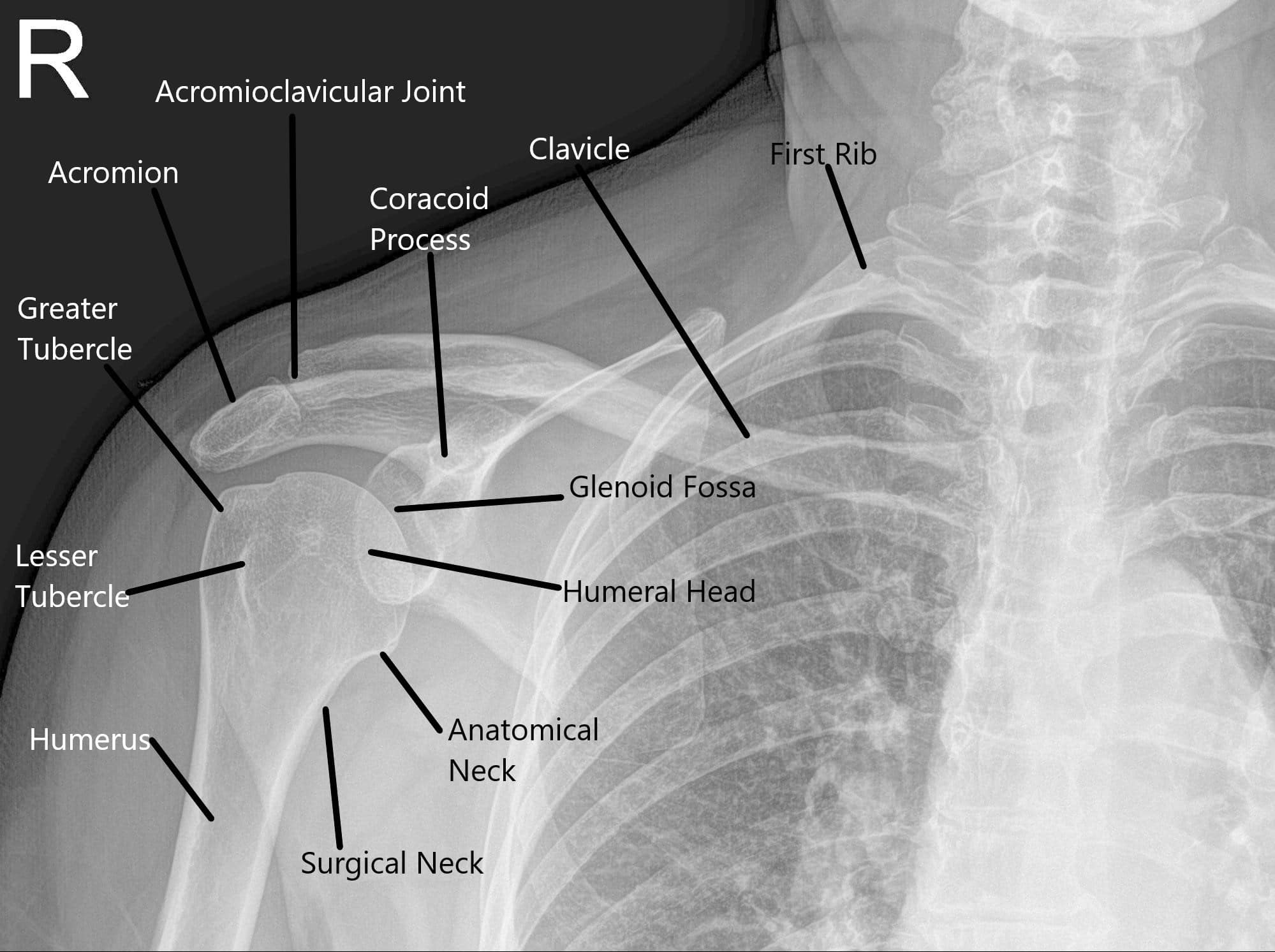
Gender: Male. Annotated image. Anatomy at the shoulder joint. The scapula articulates with the humerus and clavicle at the glenohumeral joint and coracoclavicular joints.

shoulder xray anatomy
Fig. 3.1. Anteroposterior shoulder radiograph. While achieving anteroposterior shoulder X-ray in neutral position, the patient is erect or in supine position. Central X-ray should be directed to 2.5 cm inferior to the coracoid process. Anteroposterior shoulder view allows assessment of especially the humeral head lesions and clavicular fractures.

Xray Vision Shoulders and Elbows — Taming the SRU
Typical X-ray findings in posterior shoulder dislocation include: AP view: the glenohumeral joint will be widened and the humeral head will take on a classic "light bulb" appearance due to forced internal rotation of the humerus. Lateral view: the humeral head will lie posterior to the glenoid fossa. Figure 5.

Scapula Anatomy Xray
Shoulder: annotated projections.

Shoulder Radiology Musculoskeletal Key
The shoulder, or shoulder joint, is the connection between the upper arm and the thorax. Comprising numerous ligamentous and muscular structures, the only actual bony articulations are the glenohumeral joint and the acromioclavicular joint (ACJ). The shoulder allows for an extensive range of motion due to the spheroid shape of the glenohumeral.
Normal Shoulder X Ray Left slidesharedocs
This is a basic article for medical students and other non-radiologists. A shoulder series (or shoulder x-ray) is most frequently performed following trauma looking for evidence of fracture or dislocation.. Reference article. This is a summary article.For more information, you can read a more in-depth reference article: shoulder series. Summary

Pin by Stelios Daskalogiannis on ΩΜΟΣ Medical anatomy, Medical
Posterior shoulder dislocation. less than 5% of glenohumeral dislocations but often overlooked. common in adults following a seizure or in the elderly. humeral head forced posteriorly in internal rotation whilst arm is abducted. classically, the humeral head is rounded on AP - light bulb sign. associated with anteromedial fracture of humeral head.

Pin on Anatomy Imaging
glenoid version for total shoulder arthroplasty. Magnetic Resonance Imaging. Overview. MRI is best for evaluating soft tissue structures and evaluating bone contusions or trabelcular microfractures. the stronger the magnet, the higher the intrinsic signal-to-noise ratio (e.g. a 3 Tesla MRI machine has 9x the proton energy of a 1.5 Tesla MRI.
Snapping Shoulder Causes & Management Complete Orthopedics
Description. Labeled Shoulder X-Ray Anatomy by Dr. Naveen Sharma - theRadiologist @radiologistpage #Shoulder #XRay #Anatomy #clinical #radiology #labeled #msk #diagnosis.
